The Visual Pathways |
|
| The
Retina |
- Receptors: Rodes and cones
- First order neurons: bipolar cells
- Second order neurons: ganglion cells
|
 |
| |
|
| The
Optic Nerve and Optic Chiasma |
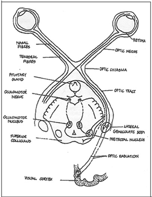 |
- Axons of ganglion cells form the optic nerve, optic chiasma and optic tracts
- Fibres from the nasal
side (with information from the temporal
visual field) remain medial and decussate at the optic chiasma
- Fibres from
the temporal side (with information from the nasal visual field) remain lateral and ipsilateral.
- Optic tracts projects predominantly to lateral geniculate body but also to:
a) Pre-tectal areas
b) Superior colliculus
c) Hypothalamus
- Third order neurons project mainly to the lingual and cuneus gyri around the calcarine sulcus.
- A few project to the superior temporal gyrus
|
 |
|
| |
|
|
Visual
Reflexes |
|
| Involuntary Eye Movements |
| Reflex
turning of the eyes |
| This is towards an object without
being conscious of it:
- Afferent
arc: Three neurons from the retinal ganglion cells to
the superficial layers of superior colliculus, then to the deep layers.
Tectobulbar neurons to the nuclei of cranial nerves III, IV, and VI
(actually to the medial reticular formation adjacent to cranial nerve
nuclei III, IV and VI)
- Efferent
arc: One neuron from the cranial nerve nuclei to the
extra-ocular muscles.
In simple eye movements, there is often some accompanying
turning of the head and neck, which is accomplished by collicular neurons
that project to anterior horn cells of the cervical spinal cord via
the tectospinal
neurons. |
| |
|
| The
Accommodation - Convergence reflex |
This reflex involves three
changes:
- Convergence of the eyes due to the action of the
medial recti muscles.
- Increased convexity of the lens.
- Pupillary constriction.
Afferent
arc: Retino - LGN - striate projection. Efferent
arc: Corticofugal projection to the
superior colliculus, and from this visual centre connections to:
- The oculomotor nuclei (for ocular convergence)
- The nucleus of Edinger-Westphal (for pupillary constriction
and increased lens convexity). This reflex is spared in Argyll-Robertson
pupil.
|
| |
|
| Fixation
reflex : |
| This for example, occurs when both
eyes follow the trajectory of an object in the smooth rather than jerky
fashion thus ensuring that any point on an object is projected onto
corresponding points of the two retinas.
- Afferent
arc: Two neurons: from the retinal
ganglion cells to the LGN, from LGN to the striate cortex.
- Efferent
arc: From the striate cortex to
the superior colliculus and from the superior colliculus to the extra-ocular
muscles via the rectobulbar
pathways.
|
| |
| |
|
| Voluntary
eye movements |
- Afferent
arc: From the retinal ganglion cells to the LGN - from
the LGN to the striate cortex via the geniculostriate pathway. From
the striate cortex to association visual cortex then to the
frontal eye field, which is located
in the caudal part of the middle frontal gyrus.
- Efferent
arc: Corticobulbar projections, ending
within the superior colliculus. The superior colliculus makes connection
via tectobulbar pathway with cranial nerve nuclei III, IV and VI.
The final projection is to the extra-ocular muscles. This pathway
mediates conjugate eye movements of which the individual is aware,
such as movements of command elicited by instructing a patient to
look to the left or to the right.
|
| |
|
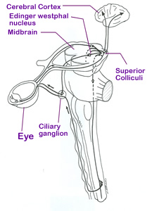
| Pupillary
light reflex |
| This is the reflex constriction of
the pupil due to increased light intensity:
- Afferent
arc: From the retina to the pretectal area, and from
thence bilaterally to the nuclei of Edinger-Westphal.
- Efferent
arc: Two neurons (parasympathetic)
from the nucleus of Edinger-Westphal to the ciliary ganglion and from
thence to the pupillary muscles of the eye.
Pupillary dilatation
- Pupillary dilation which can occur in
response to a sudden decrease in light intensity, to severe pain or to
certain emotional conditions, is due to a different pathway.
- The pathway
leads from the thalamus to the hypothalamus, and then to the intermediolateral
cell column of the upper thoracic cord through relay in the reticular
formation.
- Two neurons pass, to pupillary dilator muscles via the superior
cervical sympathetic ganglion.
|
 |
| |
|
| The Argyll-Robertson
pupil |
| This is a disorder in which the pupils
are non-reactive to light shined to the retina. The commonest cause is
neurosyphilic affliction of the pretectal nuclei or periaqueductal region,
ciliary ganglion, singly or in combination. |
| |
|
| |
|
| The
Accomodation-Convergence: |
This reflex involves three changes:
- Convergence of the eyes due to the action of the
medial recti muscles.
- Increased convexity of the lens.
- Pupillary constriction.
Afferent
arc: Retino - LGN - striate projection. Efferent
arc: Corticofugal projection to the superior colliculus,
and from this visual centre connections to:
- The oculomotor
nuclei (for ocular convergence) and
- The nucleus of Edinger-Westphal (for pupillary constriction
and increased lens convexity). This reflex is spared in Argyll-Robertson
pupil.
|
| |
|