|
The Rectus Sheath |
|
Location
- Fibrous compartment for rectus abdominis muscle in the paramedian abdominal wall.s
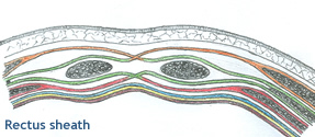
Formation
- Formed of the aponeurosis of abdominal
muscles.
- It has a posterior layer and anterior layer.
Proximal 1/3rd
- The anterior layer joins
the aponeurosis of the external oblique to form the anterior wall
of the rectus sheath.
- The posterior layer
joins with the aponeurosis of the transversus abdominis to form the
posterior wall of the rectus sheath.
Middle 1/3 rd
- Aponeurosis of internal oblique joins external oblique aponeurosis to form anterior wall.
- Posterior wall is formed by aponeurosis of transversus abdominis muscle
Distal 1/3 rd
- Mid way between umbilicus and pubic crest all three aponeurosis form the anterior layer
- The posterior layer is formed only by fascia transversalis
Note:
  |
- The anterior and posterior layers fuse in the midline
to form the linear alba, a fibrous intersection
extending from the xiphoid process to the pubic symphysis.
- The inferior ¼ of the rectus sheath is deficient
posteriorly. The limit of the posterior wall is marked by the arcuate
line
- The lateral margin of rectus sheath is called linea semilunaris
|
|
|
| |
|
Contents of Rectus Sheath |
- Rectus abdominis muscle
- Inferior and superior epigastric vessels
- Terminal parts of the lower five intercostal nerves, and the Subcostal nerve.
- Fibro fatty connective tissue
- Occasionally lymph node(s)
|
| |
|
Extra peritoneal fascia |
- Transparent membrane' which lines the
inside of the abdominal wall.
- Its parts are named according to what
it lines e.g.
(+)
diaphragmatic fascia;
(+) iliac fascia;
(+) psoas fascia.
(+) fascia transversalis ( part covering
the muscle transversus abdominis ).
|
| |
|