Home | OG anatomy | Gross Anatomy | Topic index | Chapter 22
| contents | |
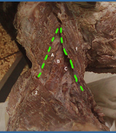
The external jugular vein |
|
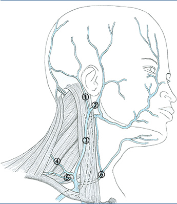 |
• This vein is formed near the angle of the mandible by the union of the posterior branch of retromandibular and posterior auricular veins . • It crosses sternocleidomastoid muscle, runsover the roof of the triangle and joins the subclavian veins • The vein drains most of the scalp and face on the same side. • This vein dilates and becomes visible in fluid overload, in heart failure in SVC obstruction, prolonged raised intrathoracic pressure, e.t.c. • The walls of the vein are attached to the deep fascia. If the vein is lacerated, the fascia pulls the vein open and blleding is severe. Also, air embolism could follow.
|
| Arteries | |
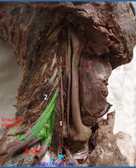 |
• The subclavian artery (third part) -B • Transverse cervical artery from the thyrocervical trunk to supply muscles in the scapular region. • Suprascapular artery from the thyrocervical trunk. • Occipital artery, from the external carotid artery. Note the boundaries: A: Anterior scalene muscle |
| Nerves | |
 |
• Spinal accessory nerve to the sternocleidomastoid muscle and the trapezius muscle. • Cervical plexus and its cutaneous branches from up downwards. • Lesser occipital nerve (c2) • Great auricular nerve (c2 c3) • Transverse cervical nerve (c2 c3) • Suprascapular nerves (c3c4) Supraclavicular part of the brachial plexus |
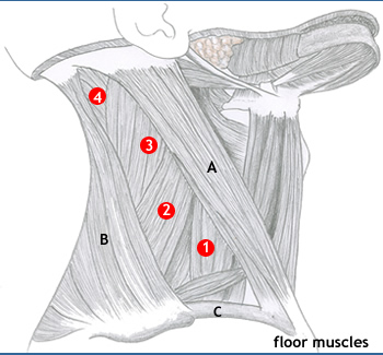
| Muscles | |
|
|
| subdivisions | |
|
|