The Midbrain

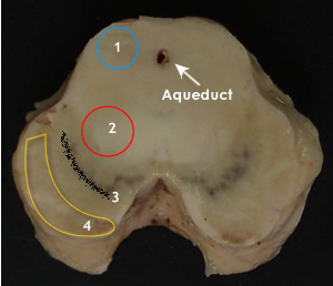
The central aqueduct is distinctive. Dorsal to this is the tectum with four colliculi.

  • Superior colliculi process visual sensations.
  • Inferior colliculi process auditory sensations.

Red nucleus is most distinctive in the midbrain tegmentum. It integrates information from cerebrum and cerebellum (involuntary motor control)

Its fibers to the spinal cord (rubrospinal) transmit impulses that facilitate flexor muscle tone.

Structures dorsal and lateral to the red nucleus form the midbrain reticular formation.

What do you consider to be the main functions of the reticular formation?


The mibrain consists of four main parts:

  • Tectum(1)
  • Red nucleus(2)
  • Substantia nigra (3)
  • Crus cerebri (4)

Around the aqueduct is the periaqueductal gray. It contains nuclei of oculomotor and trochlear nerves.

The most prominent structure in the tegmentum is the red nucleus but the tegmentum, also contains ascending tracts.

The substantia nigra consits of melanin containing neurons tha regulate motor output of cerebral cortex

Crus cerebri represents a bundle of projection fibers.


The following structures can be identified In a cross section of the midbrain.

  • Mesencephalic nucleus.
  • Medial lemniscus.
  • Decussation of superior cerebellar peduncle.
  • Inferior/superior Colliculi.
  • Descending fibers from motor cortex.
  • Nuclei of cranial nerve and gray matter.
  • Periaqueductal gray
  • Oculomotor/trochlear nucleus
  • Edinger Westphal
  • Red nucleus
  • Substantia nigra
  • Mesencephalic reticular formation

Blood supply to the midbrain
Most of the blood supply is derived from branches of the basilar artery. 

  1. Posterior cerebral
  2. Superior cerebellar
  3. Posterior communicating
  4. Anterior choroidal

Vascular lesions of the midbrain include:
Weber‘s syndrome, which involves the basal portion of the midbrain due to occlusion of a mesencephalic branch of the posterior cerebral artery.


Area of infarction involves:

  1. Pyramidal fibres- contralateral hemiparesis. 
  2. Oculomotor nerve - ipslateral paralysis of the ocular muscles except the lateral rectus and superior oblique.

Red area: shows area of vascular lesion involvement


Click on hot spots to show names