Medulla Oblongata


The medulla extends from foramen magnum to pons.

Transition from spinal cord to medulla is characterized by:

  • Obliteration of median fissure by pyramidal decussation
  • Appearance of gracile and cuneate tubercles
  • Appearance of cranial nerves
  • Appearance of fourth ventricle
  • Dissapearance of spinal nerves
  • Cranial nerves associated with the medulla include:
    • Hypoglossal - between pyramid and olive.
    • Glossopharyngeal post -olivary sulcus.
    • Vagus post - olivary sulcus.
    • Accessory post - olivary sulcus.
    • Vestibulocochlear - cerebellopontine angle.

The caudal medulla is closed but the cranial half is open dorsally (fourth ventricle).


Its external appearance is characterized by the pyramid anteriorly, olive anterolaterally, tuberculum cinereum posterolaterally , gracile and cuneate tubercles dorsally and the attached nerves

The medulla relays sensory information to thalamus and contains major regulatory centers in its reticular formation

It also a number of nuclei:

  • Relay nuclei - gracilis, cuneatus, olivary.
  • Sensory and motor nuclei of cranial nerves vestibulocochlear, glossopharyngeal, vagus, accessory and hypoglossal nerves).
 

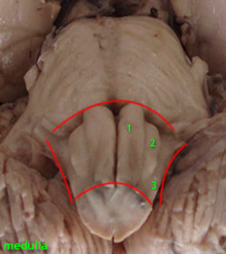
The internal anatomy of the closed and open medulla demonstrates the following structures .

  • Fourth ventricle and central canal and central gray
  • Nuclei:
    • Hypoglossal
    • Cuneate and accessory cuneate
    • Gracile
    • Trigeminal (spinal)
    • Ambiguus (for IXN, XN, XIN)
    • Dorsal motor nucleus of vagus
    • Vestibular
    • Olivary
    • Reticular
 

Fiber bundles  
  • Fasciculus gracilis
  • Fasciculus cuneatus
  • Pyramidal
  • Spinal tract of trigeminal
  • Medial lemniscus
  • Internal arcuate
  • Hypoglossal fibers
  • Inferior cerebellar peduncle
  • MLF

Blood supply to the medulla oblongata


Four main arteries supply the medulla oblangata

•  Anterior spinal arteries supplying the antero-medial structures namely

- Pyramids

- Medial lemniscus

- Hypoglossal nucleus

- Medial Longitudinal fasciculus

- Solitary and vagal nuclei

•  Posterior spinal arteries supplying the fasciculus and nucleus gracilis and cuneatus

•  Posterior inferior cerebellar artery supplying the retro-olivary region, including

- Spinothalamic tract

- Spinal trigeminal nucleus

- Nucleus ambiguus

Hypothalamo spinal tracts

•  Bulbar branches of the vertebral artery supplying the pyramids, hypoglossal, nucleus, and inferior olivary nuclear complex.


Tracts of the Medulla Oblangata

The medulla oblangata contains all the tracts ascending to the brain, and those descending to the spinal cord.  Key among them are


Ascending tracts   
Descending tracts
  • Medial lemniscus
  • Fasciculus cuneatus
  • Fasciculus gracilis 
  •  Spinocerebelar tracts
  • spinotectal tracts
  • ventral spinothalamic
  • Rubrospinal
  • Vestibulospinal
  • Reticulospina
  • lSpinal tracts of the trigeminal
  • Cortico-spinal tract

Nuclei of the Medulla Oblangata

Nucleus

Function

Main connections

Solitarius (Gustatory)

Taste

Thalamus, CN VII, IX, X

Gracilis and cuneatus

Proprioception and fine touch

Fasciculus gracilis and cuneatus, Thalamus, Cerebellum

Inferior olivary

Control of muscle Activity

Spinal cord, Cerebellum

Inferior salivatory

Control of parotid gland

Gustatory nucleus ,CN IX

Reticular formation: vasomotorand respiratory centres. Chemoreceptor trigger zone

Control of ardiorespiratory and  GIT functions.Control of motor activity

Vagal nuclei, nuclei of phrenic and intercostals nerves. Spinal cord. Brain stem.


Applied anatomy

Medial medullary syndrome results occlusion of a anterior spinal artery or bulbar branches of the vertebral artery.

Structures normally included in the area of infarction are:

i) Medial lemniscus

ii) Pyramid

iii) Hypoglossal nerve

Sensations of position, movement, discriminative touch and vibrations on the opposite side of the body (interruption of medial lemniscus) and ipsilateral paralysis of the tongue due to the involvement of the hypoglossal nerve.

This disorder forms an example of a crossed or alternating paralysis in which the body is affected on the opposite side, but the muscles supplied by the cranial nerve affected are on the same side. This particular condition is known as “ alternating hypoglossal hemiplegia ” or inferior alternating hemiplegia .

Lateral medullary syndrome

Results from occlusion of the posterior inferior cerebellar artery or a medullary branch of it.

Lesions normally includes the following structures with the respective deficits:

i) Spinal trigeminal tract and nucleus : Ipsilateral loss of pain and temperature in the areas of distribution of the trigeminal nerve.

ii) Spinal lemniscus : loss of pain and temperature sensibilities on the opposite side of the body. Touch is diminished rather than lost (explain why).

iii) Nucleus ambiguus: ipslateral paralysis of the muscle of the palate in swallowing and phonation (explain why).

iv) Hypothalamospinal tract : Horner's syndrome characterised by small pupil, ptosis (drooping of eyelids) and anhidrosis (explain why).

Vi)  Inferior cerebellar penduncle and vestibular nuclei : if included caused dizziness, cerebellar ataxia and nystagmus.