The Urinary bladder |
|
|
|
| Location |
|
 |
- In the adult, the bladder lies in the minor pelvis, posterior to the pubis.
- It is separated by the retropubic space with fat.
- In infants and children, the bladder is in the abdomen, beginning to enter the pelvis major at about 6yrs.
- It reaches the pelvis at puberty.
- The shape, size, positions and relations vary with the amount of urine it contains and with age.
|
|
Peritoneal relations |
|
|
 |
- The superior part is covered posteriorly, cranially and anteriorly
- vesico uterine pouch in the females and the recto vesical pouch in the male.

|
|
| Relations |
|
|
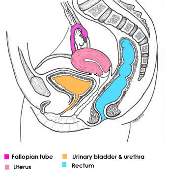
Female |
Male |
|
Anteriorly |
- Levator ani
- Obturator internus
|
- Pad of fat
- Levator ani
- Obturator internus
|
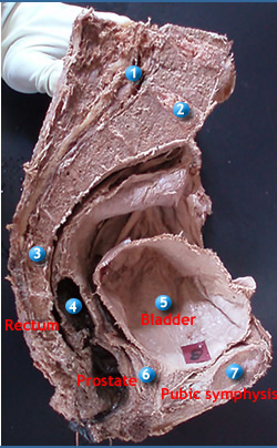
Posteriorly |
|
- Vas deferens
- Seminal vesicles
- Rectum
|
Superiorly |
- Coils of small intestine (when full)
- Body of uterus (when empty)
|
|
Inferiorly |
- Pelvic
- Urogenital diaphragm
|
- Urogenital diaphragm
- Pelvic diaphragm
- Prostate surrounds the urethra
|
|
|
| Surfaces |
|
- Superior surface, facing upwards
- Two inferior lateral surfaces, facing inferiorly
- Posterior surface facing posteriorly and inferiorly. This is also called the base
- Where the base and infero lateral surfaces converge, is called the neck of bladder.
- This is where the lumen of the bladder opens into the urethra and rests on the prostate gland in the male.
|
| support |
|
|
- The entire organ is enveloped by vesical fascia, a layer of loose connective tissue in which there is the vesical venous plexus
- The neck is held firmly by pubo prostatic ligaments in males and the pubo vesical ligament in the female.
- Major support is derived from the pelvic and urogenital diaphragms.
|
|
|
Blood supply |
|
|
- Superior vesical arteries from the umbilical artery
- Inferior vesical arteries from the internal iliac
- Small branches from obturator and inferior gluteal arteries
- In females, uterine and vaginal arteries give branches to the bladder.
- The veins correspond to the arteries. From the vesical venous plexus and are tributaries of the internal iliac vein.
- Note: The vesical venous plexus in males envelopes the base of the bladder and prostate. The seminal vesicles, ductus deferens and inferior ends of the ureters
- It is connected to the prostatic venous plexus
- It may drain via sacral veins into the vertebral venous plexus
- In females it envelopes the pelvic part of the urethra and the bladder neck. It receives blood from the dorsal vein of the clitoris.
- It communicates with the vaginal plexus
|
|
Lymphatics |
|
|
- From the superior part – external iliac nodes
- From the inferior part- internal iliac nodes
- Some from the neck- sacral and / or common iliac nodes
|
|
Innervation |
|
|
- Parasympathetic fibres from the pelvic splanchnic nerves are motor to the detrusor muscle and inhibitory to the internal sphincter of the bladder
- Sympathetic fibres are derived form T11- L2.
- These fibres are inhibitory to the bladder and excitatory to the internal sphincter muscles.
- The nerves form a vesical plexus consisting of both sympathetic and parasympathetic fibres. It is continuous with the inferior hypogastric plexus.
- The external sphincter with other perineal muscles is supplied by pudendal nerve.
|
|