SUMMARY OF THE ORGANIZATION OF THE PERINEUM |
|
| Boundaries |
|
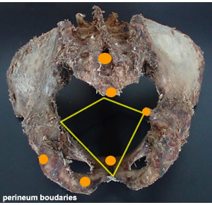 |
- The Pubic Symphsis
- The Inferior pubic rami
- The Ischial rami
- The Ischial tuberosities
- Sacrotuberous ligaments
- The coccyx
|
|
| Divisions |
|
|
 |
The line joining the two ischial tuberosities divides the perineum into two triangular parts
- The anal triangle (A)
Posterior to this line
contains the anus
- The urogenital region (B) ,
Anterior to this line
Contains the scrotum
Root of the penis (or the vulva in the female)
|
|
| The Pelvic Diaphragm |
|
Components
- Lavetor ani
- Coccygeous muscles
|
|
|
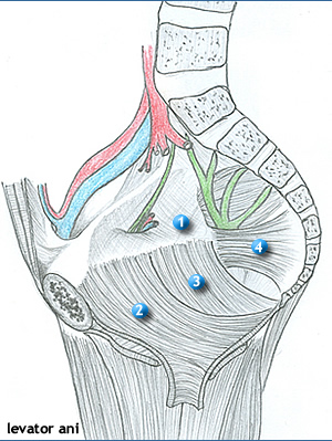
| Levator Ani |
|

|
|
|
Attachments of the lavetor ani |
|
Origins
- The pelvic surface of the body of the pubis
- Tendinous arch, formed by a thickening of the parietal pelvic fascia over the obturator internus muscle.
- Ischial spine
Insertions
- Central perineal tendon
- The wall of the anal canal
- Anococcygeal ligament
- Coccyx
Parts of Levator Ani
Pubococcygeous |
Puborectalis |
Lavetor prostatae |
|
| The main part.
- Arises from the pubis
- Inserts into the coccyx, and the Anococcygeal ligament.
It is located between the anal canal and the tip of the coccyx
encircles the:
a) Urethra
b) Vagina
c) Anus
- Merges into the central perineal tendon.
|
- Part that loops around the anorectal junction
- Arises from the pubis.
|
- Anterior fibres that run inferior to the prostate.
- In the females, these fibres constitute the pubo vaginalis. |
|
| Functions of the lavetor ani |
- Constitutes the pelvic floor for the containment/support of the pelvic viscera – resisting inferior thrust
during e.g. coughing, deep expiration e.t.c.
- They raise the pelvic floor, assisting the anterior abdominal wall muscles in compressing the viscera e.g. in
coughing, deep expiration, vomiting, urinating
- Support of the prostate and the vagina providing sphincteric action sto the latter, and urethra
- The puborectalis holds the anorectal junction anteriorly, increasing angulation between the rectum, and
anal canal. This prevents passage of feces from the rectum into the anal canal when defecation is not
desired, or is inconvenient.
- The gutter arrangement facilitates the rotation of the baby’s head during parturition, in accordance with the pelvic diameters.
- Supports the fetal head (while cervix is dilating during parturition)
|
|
The coccygeus: |
|
|
- Continuous with the iliococcygeous muscle
- Forms the posterior and smaller part of the pelvic diaphragm.
- Arises from the pelvic surface of the ischial spine and sacrospinous ligament
- Inserts on the lateral margins of the 5th sacral vertebra and coccyx.
- The fibres support the coccyx, and pull it anteriorly, after it has been pressed posteriorly during child-birth
|
|
| Structures piercing the pelvic diaphragm |
|
- Anus
- Urethra
- Vagina (in females
|
|