The Middle Mediastinum
|
|
| The pericardium:
|
|
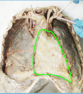 |
 Double walled fibroserous sac Double walled fibroserous sac
Encloses:
- Heart
- Roots of pulmonary trunk and aorta
- Termination
of pulmonary veins and vena cavae.
Location:
- Middle mediastinum
- Posterior to the body of the sternum and th 2 nd to 6 th costal
cartilages
- Anterior to T5-8 vertebrae.
|
|
|
| Relations |
|
Superior:Pierced by the
- Aorta,
- pulmonary trunk
- Superior vena cavae.
- In children it is related to the thymus
Inferiorly:
- Separated from the liver and fundus of the stomach by the central tendon
of the diaphragm with which it fuses.
Anteriorly:
- Seperated from the body of sternum and 2 nd to 6 th costal cartilages
by pleura with the lungs except at the cardiac notch.
- The internal thoracic
vessels and parasternal nodes run along the lateral borders of the sternum.
- In the midline it is attached to the sternum by sternopericardial ligaments
Posteriorly:
- The contents of the posterior mediastinum
Laterally:
- The
phrenic nerve
- Periocardiophrenic vessels
- Mediastinal surface
of the lungs and their covering visceral pleura( what structures are
found at the hilum of the lungs?)
|
| |
|
|
Organization of the pericardium
|
|
| Consists of two layers:
- The fibrous - tough aponeurosis-like
outer indistensible jacket
- The serous - moist inner
lining with two layers
(+) Parietal layer lining the inside of
the fibrous pericardium
(+) Visceral pericardium intimately lining
the heart surface.
The space between the visceral and parietal pericardium
is called pericardial cavity. It contains a thin film of fluid allowing frictionless movement of the heart |
| |
|
|