| THE
CARPAL TUNNEL |
|
Boundaries: |
|
| Anterior: |
 |
Flexor retinaculum
|
|
Posterior: |
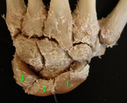 |
Carpal bones (lunate, triquetral, scaphoid, trapezoid) |
|
Medial: |
| Hook of hamate, pisiform |
|
Lateral: |
| Tubercle of scaphoid, ridge of trapezium |
|
Contents: |


|
|
| |
|
| |
Note:
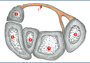
- The four tendons of the superficial flexor are separate and lie in two rows, with the middle and ring finger tendons in front of the index and little finger tendons.
- The tendons of flexor digitorum profundus lie deeply in one plane.
- All the eight tendons (of superficialis and profundus) share a common flexor sheath.
- The tendons of flexor policis longus lies in its own synovial sheath.
- At the lateral end of the tunnel a deep lamina from the flexor retinaculum is attached to the medial lip of the groove on the trapezium.
- The tendon of flexor carpi radialis, enclosed in its own synovial sheath, runs in the groove in this subcompartment of the carpal tunnel.
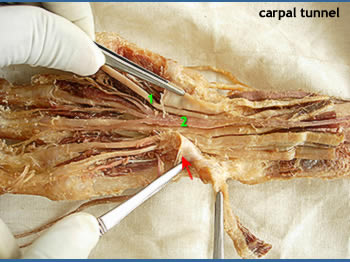
- The median nerve passes deep the flexor retinaculum between the flexor digitorum superficialis tendon to the middle finger and the flexor carpi radialis.
|
|
Carpal tunnel syndrome: |
|
 Compression
of the median nerve in the carpal tunnel due to arthritic changes in
the wrist joint, synovial sheath thickening or edema. Symptoms: Compression
of the median nerve in the carpal tunnel due to arthritic changes in
the wrist joint, synovial sheath thickening or edema. Symptoms:
- Impaired sensation over three and half digits on the thumb side
- Wasting and weakening of thenar muscles
|
| |