The Leg And Dorsum Of The Foot

Dorsum of Foot


Cutaneuos nerves  

Medial margin : Saphenous nerve

Lateral margin:  Sural nerve

Dorsum Centre:  Superficial peroneal

1st Interdigital cleft:  Deep peroneal


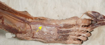
Dorsalis Pedis artery


Origin:

Continuation of anterior tibial artery


Course:

  • At the base of first inter-metatarsal space

  • Lateral to tendon of extensor hallucis longus

  • Crossed by tendon of extensor hallucis brevis

  • Palpable lateral to EHL on a line from midpoint between the two malleoli and first toe cleft.

Branches

Lateral tarsal artery: Runs beneath extensor digitorum brevis

Arcuate artery: Beneath extensor digitorum brevis over bases of metatarsals

1st dorsal metatarsal artery

Termination and anastomosis

  • Enters plantar foot through 1st inter-metatarsal space

  • Joins lateral plantar artery to complete plantar arch

Distribution

  • Soft tissues on dorsum of foot
  • Tarsals, metatarsals and phalanges
  • Plantar aspect of foot


Dorsal venous arch  

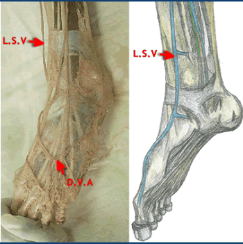
Lies over the heads of metatarsals
Receives most of its blood from:

  1. Marginal and interossseous tributaries from the sole of the foot
  2. Dorsal digital veins

Joins medial dorsal digital vein medially to form great saphenous vein

Laterally drains into small saphenous vein

(Dorsal Venous arc [D.V.A] forming the long saphenous vein [L.S.V] on the medial side of the foot )


Inferior extensor retinaculum  
  • Y –shaped thickening of deep fascia

  • Arises from upper surface of calcaneus

  • Upper limb attaches to medial malleolus

  • Lower limb blends with plantar aponeurosis

  • Prevents bow-stringing of the extensor tendons as they pass across the ankle joint.


Inferior Extensor Retinaculum (IER)

Superior Extensor Retinaculum (SER)

Identify the structures labelled A, B, C.